Visualizing Mitosis Poster Manufacturer,Supplier and Exporter in India

Product Code : EL-CS-10777
Quick Answer: The Visualizing Mitosis Poster shows detailed illustrations of each mitotic stage beside close-up photomicrographs of an onion root tip and a whitefish blastula, and measures 890 x 585 mm.
Last updated: October 2026
Key Features
- Detailed illustrations of each mitotic stage
- Close-up photomicrographs of an onion root tip undergoing mitosis
- Close-up photomicrographs of a whitefish blastula undergoing mitosis
- Lets students compare plant and animal mitosis
- Dimensions of 890 mm x 585 mm (w x h)
Technical Specifications
| Specification | Value |
|---|---|
| Width | 890 mm |
| Height | 585 mm |
| Content | Illustrations of each mitotic stage with photomicrographs |
About This Product
This poster sets drawings of each stage of mitosis next to matching photomicrographs, using an onion root tip for plants and a whitefish blastula for animals. Students can use it to review the steps of mitosis and compare how they take place in plant and animal cells.
Frequently Asked Questions
What size is the Visualizing Mitosis Poster?
It measures 890 mm wide by 585 mm high.
Which specimens are shown?
Photomicrographs of an onion root tip and a whitefish blastula undergoing mitosis.
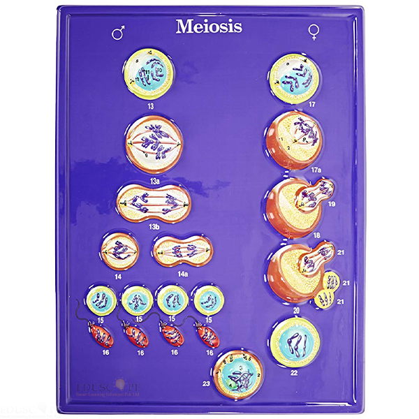
Related: Cell Slides, Meiosis Model and Osmosis Simulation Activity Model.
For a quotation on the Visualizing Mitosis Poster, contact Didactic Lab.