Meiosis Microslide Manufacturer,Supplier and Exporter in India
Product Code : EL-CS-10772
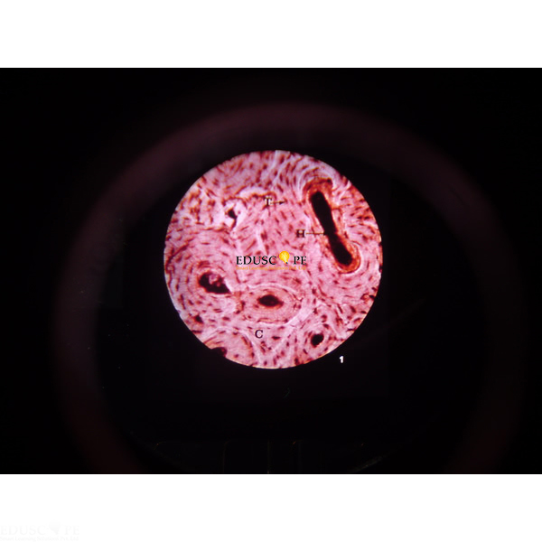
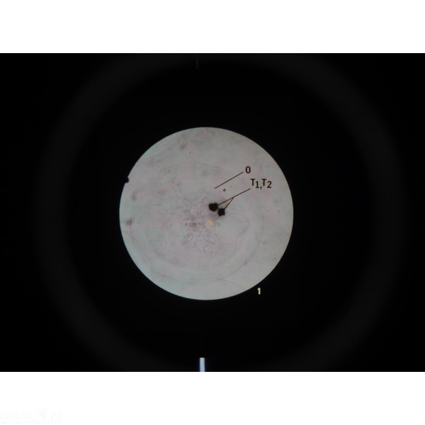
Quick Answer: The Meiosis Microslide is a set of 8 related 35 mm photomicrograph images that follow the egg from primary oocyte stages to a zygote ready for first cleavage.
Last updated: October 2026
Key Features
- 8 related 35 mm images photographed through a microscope
- Stages from primary oocyte with tetrads formed to zygote ready for first cleavage
- Four of the images taken at 750x
- Arrows and callouts mark the features being studied
- Text folder with a pocket for storing the Microslides
Technical Specifications
| Specification | Value |
|---|---|
| Images | 8 related 35 mm images |
| Viewing | Viewer |
| Magnification noted | 750x on four images |
| Supplied with | Text folder with storage pocket |
About This Product
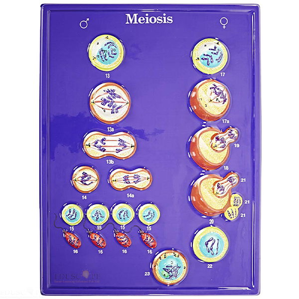
The images show a primary oocyte with tetrads formed, with dyads separated, with tetrads on the spindle and with the first polar body forming, then a secondary oocyte with chromosomes formed, the pronucleus stage, a mature egg and a zygote ready for first cleavage. Four of the images are at 750x. A detailed text folder goes with the set to inform students and question them on the topic. Export enquiries for the Meiosis Microslide are welcome from Lebanon, Zimbabwe, Venezuela, Djibouti, Afghanistan and Saint Lucia.
Frequently Asked Questions
Which stages does the Meiosis Microslide show?
Primary oocyte stages (tetrads formed, dyads separated, tetrads on spindle, first polar body forming), secondary oocyte, pronucleus stage, mature egg and zygote ready for first cleavage.
How are the images viewed?
The 8 images are 35 mm and are viewed through a Viewer.
Related: Cell Slides, Meiosis Model and Ultra Structure of Animal Cells Microslide.
For a quotation on the Meiosis Microslide, contact Didactic Lab.