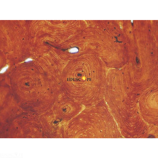
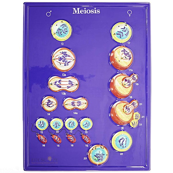
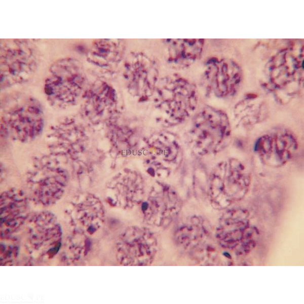
Ultra Structure of Animal Cells Microslide Manufacturer,Supplier and Exporter in India
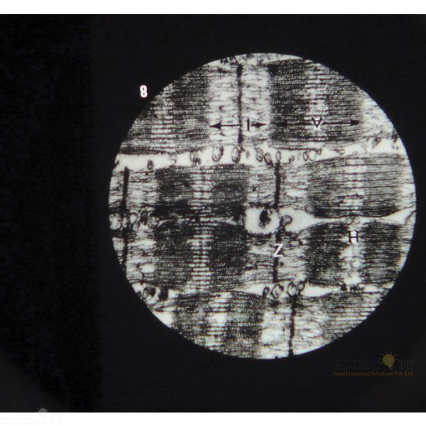
Product Code : EL-CS-10773
Quick Answer: The Ultra Structure of Animal Cells Microslide is a set of 8 related 35 mm images of cell structures such as the mitochondrion, centriole and Golgi body, photographed through a microscope.
Last updated: October 2026
Key Features
- 8 related 35 mm images photographed through a microscope
- Covers nerve cell membrane, gland cells and plasma cell
- Shows Golgi body and vacuole, mitochondrion and centriole
- Includes lamp-brush chromosomes and muscle cell fibrils
- Arrows and callouts, with a text folder that has a storage pocket
Technical Specifications
| Specification | Value |
|---|---|
| Images | 8 related 35 mm images |
| Viewing | Viewer |
| Subjects | Nerve cell membrane, gland cells, plasma cell, Golgi body and vacuole, mitochondrion, centriole, lamp-brush chromosomes, muscle cell fibrils |
About This Product
This Microslide looks inside animal cells at the level of individual structures, from the nerve cell membrane and plasma cell to lamp-brush chromosomes and muscle cell fibrils. Each image carries arrows and callouts so students can find the features under study. The set is viewed in a Viewer and comes with a detailed text folder that asks students questions on the topic.
Frequently Asked Questions
What does the Ultra Structure of Animal Cells Microslide show?
Nerve cell membrane, gland cells, plasma cell, Golgi body and vacuole, mitochondrion, centriole, lamp-brush chromosomes and muscle cell fibrils.
Related: Cell Slides, Animal Cell Model and Meiosis Microslide.
For a quotation on the Ultra Structure of Animal Cells Microslide, contact Didactic Lab.