Basic Human Histology Microslide Manufacturer,Supplier and Exporter in India
Product Code : EL-CS-10774
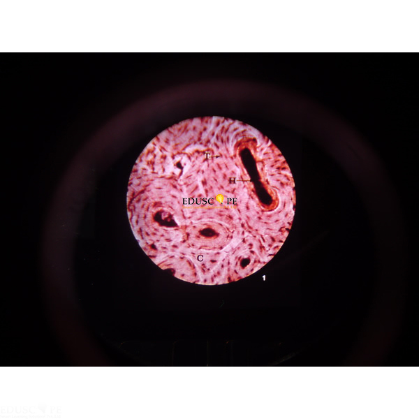
Quick Answer: The Basic Human Histology Microslide is a set of 8 related 35 mm microscope images of human tissues, from stained nerve cells and a bone cross section to red and white blood cells.
Last updated: October 2026
Key Features
- 8 related 35 mm images for a Microslide Viewer
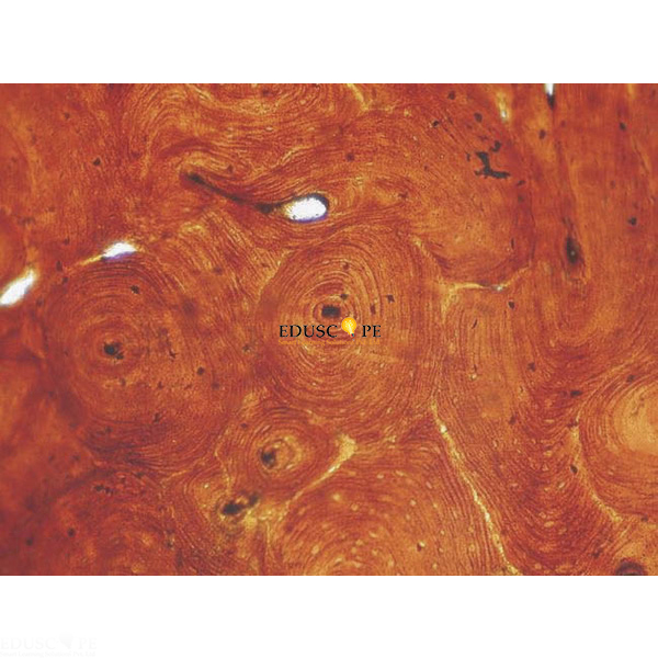
- Stained nerve cells and stained bone cross section
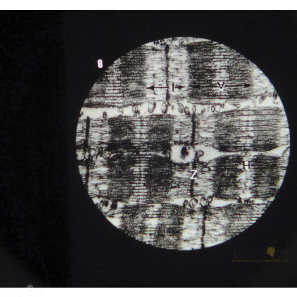
- Cartilage, striated muscle cells and fat cells
- Epithelial tissue with a ciliated epithelial cell
- Red blood cells and white blood cells
Technical Specifications
| Specification | Value |
|---|---|
| Images | 8 related 35 mm images |
| Viewing | Microslide Viewer |
| Supplied with | Detailed text folder with storage pocket |
About This Product
Eight basic human tissue types are shown in this Microslide, including cartilage, fat cells, striated muscle and epithelial tissue. Arrows and callouts guide students to the features they are studying, and the text folder that comes with it informs and questions them on the topic. The folder has a pocket to hold the Microslides. The Basic Human Histology Microslide is offered to export buyers in Chad, Niger, Chile, Papua New Guinea, Suriname and Nicaragua.
Frequently Asked Questions
Which tissues are in the Basic Human Histology Microslide?
Stained nerve cells, stained bone cross section, cartilage, striated muscle cells, fat cells, epithelial tissue with a ciliated epithelial cell, red blood cells and white blood cells.
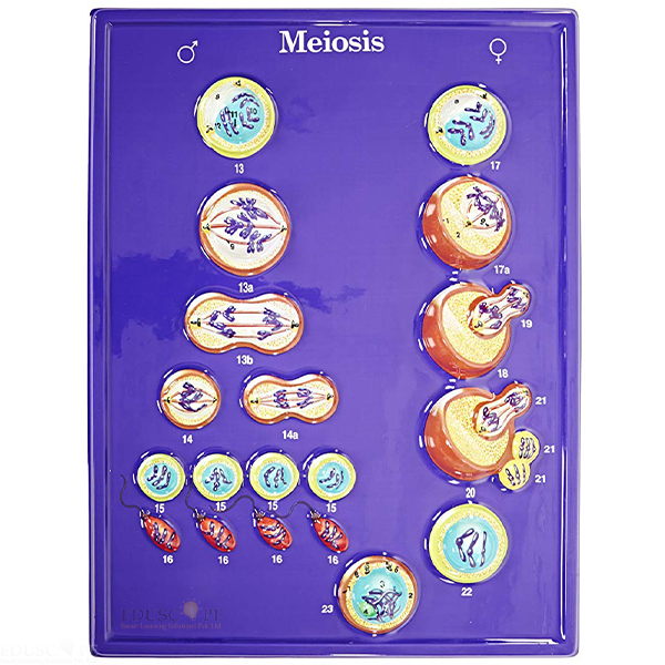
Related: Cell Slides, Basic Histology Slide Set of 25 and Meiosis Microslide.
For a quotation on the Basic Human Histology Microslide, contact Didactic Lab.